Global Specular Microscope Market Analysis: Clinical Trends, Technological Innovations, and Competitive Landscape (2026-2031)
- Single User License (1 Users) $ 3,500
- Team License (2~5 Users) $ 4,500
- Corporate License (>5 Users) $ 5,500
The global ophthalmic medical device and diagnostic sector represents one of the most technologically advanced and clinically vital segments of the modern healthcare industry. Within this highly sophisticated diagnostic landscape, the Specular Microscope market occupies a uniquely specialized and indispensable position. A specular microscope is an advanced, non-invasive, optomechanical and digital imaging device specifically engineered to visualize, capture, and quantitatively analyze the corneal endothelium—the innermost, single layer of cells on the posterior surface of the human cornea.
To understand the absolute clinical necessity of specular microscopy, one must understand the physiology of the cornea. The corneal endothelium is a fragile monolayer of primarily hexagonal cells that are derived from the neural crest during embryonic development. Crucially, human corneal endothelial cells generally do not undergo mitosis (cell division) in vivo. This means that a human is born with a finite number of these cells—typically around 4,000 cells per square millimeter (cells/mm²)—and this density naturally decreases by approximately 0.6% per year throughout life. The primary physiological function of the endothelium is to act as a metabolic fluid pump. Because the cornea is avascular (having no blood vessels) to remain perfectly transparent, it constantly absorbs nutrients and fluid from the aqueous humor inside the eye. The endothelial cells continuously pump this excess fluid back out of the stroma. If the endothelial cell density drops below a critical threshold (typically around 500 to 700 cells/mm²), the pump mechanism fails. This failure results in severe, irreversible corneal edema (swelling), profound loss of corneal transparency, debilitating pain, and ultimately, severe vision loss or blindness, a condition clinically termed bullous keratopathy.
The specular microscope functions by projecting a narrow slit of light onto the cornea at a specific angle. The light travels through the transparent anterior layers and reflects off the optical interface between the corneal endothelium and the aqueous humor, due to the slight difference in their refractive indices. The reflected light (the specular reflection) is captured by a high-resolution charge-coupled device (CCD) or complementary metal-oxide-semiconductor (CMOS) camera. Modern specular microscopes do far more than simply take a picture; they utilize highly advanced, integrated software algorithms to instantly perform automated morphological analyses. Clinicians rely on this technology to measure three critical parameters: Endothelial Cell Density (ECD), pleomorphism (the variation in cell shape, specifically measuring the percentage of ideal hexagonal cells), and polymegethism (the variation in cell size, measured as the Coefficient of Variation).
The epidemiological imperatives driving the intense demand for specular microscopes are profound and structurally permanent. The global healthcare system is facing an unprecedented wave of degenerative ocular conditions, heavily driven by a rapidly aging global population. According to comprehensive data published by the World Health Organization (WHO), an estimated 2.2 billion individuals globally exist with some form of vision impairment or blindness. Within this staggering demographic, age-related cataracts account for an estimated 94 million cases, while glaucoma affects approximately 7.6 million individuals, representing the leading causes of preventable and irreversible blindness worldwide. Furthermore, data from the Global Burden of Disease (GBD) study emphasizes the severity of this crisis, indicating that ophthalmic diseases accounted for approximately 4.5% of total global Disability-Adjusted Life Years (DALYs) in 2020.
Because standard cataract surgery (phacoemulsification) utilizes intense ultrasonic energy inside the eye, it inherently causes collateral damage and trauma to the delicate corneal endothelium, accelerating cell loss. Consequently, preoperative specular microscopy has become the absolute standard of care to identify patients with critically low cell counts or pre-existing endothelial dystrophies (such as Fuchs' Endothelial Dystrophy) before surgery, allowing surgeons to alter their surgical techniques or utilize specialized viscoelastic protective gels to prevent postoperative corneal failure. This vital risk-management utility guarantees the continuous, high-volume procurement of specular microscopes globally.
Market Scale and Growth Projections
The economic dimensions of the specular microscope market reflect its status as a mature, highly specialized capital equipment sector that is tightly integrated into the standard workflow of advanced ophthalmic surgical centers and eye banks.
• Estimated Market Size (2026): The global market for specular microscopes is projected to achieve a highly substantial valuation ranging between 127 million USD and 215 million USD by the year 2026. This robust valuation encapsulates the continuous, high-value capital procurement of advanced, fully automated, non-contact imaging platforms by major hospital networks, alongside the expanding diagnostic capabilities of independent ophthalmic clinics.
• Compound Annual Growth Rate (CAGR): Over the forecast period spanning from 2026 to 2031, the market is anticipated to expand at a steady, resilient estimated CAGR of 4.6% to 6.2%.
This stable growth trajectory is highly insulated from broader economic volatility due to the medicolegal and clinical necessity of the technology. The growth is heavily propelled by the global explosion in cataract surgical volumes, the surging popularity of premium, highly profitable intraocular lenses (IOLs) that demand pristine preoperative corneal assessments, and a vast global capital replacement cycle wherein older analog or semi-automated devices are being aggressively replaced by high-definition, AI-driven digital platforms.
Product Segmentation and Market Trends
The specular microscope market is technologically stratified by the physical modality of image acquisition and clinically segmented by the specific operational environment of the end-user. Each distinct category is experiencing specific evolutionary trends driven by computational optical advancements and shifting clinical workflows.
Classification by Type
• Non-Contact Specular Microscopes: This segment represents the absolute gold standard and commands the overwhelming majority of the global market share and revenue. Non-contact devices utilize sophisticated infrared auto-alignment and autofocus systems to capture the endothelial reflection from a distance of several millimeters away from the corneal surface, entirely eliminating any physical contact with the patient's eye.
o Technological Development Trends: The dominating trend in the non-contact segment is the aggressive optimization of patient comfort, operational speed, and diagnostic accuracy. Because nothing touches the eye, the procedure does not require topical anesthetic drops, completely eliminates the risk of transmitting highly contagious ocular pathogens (such as epidemic keratoconjunctivitis or adenoviruses), and prevents iatrogenic corneal abrasions. Modern non-contact systems are heavily integrating Artificial Intelligence (AI) and deep learning algorithms. These advanced software suites instantly and automatically trace the delicate, low-contrast cell borders in a fraction of a second, entirely removing subjective human error from the cell counting process. Furthermore, manufacturers are developing wide-field non-contact systems that can capture cells from the extreme periphery of the cornea, rather than just the central optical zone, providing a much more comprehensive map of global endothelial health.
• Contact Specular Microscopes: Representing the historical, legacy technology of the industry, contact specular microscopes operate by physically touching the cornea using a specialized applanating objective lens. The procedure requires the instillation of topical anesthetic drops and the application of a viscous optical coupling fluid to the eye.
o Technological Development Trends: While significantly losing market share to non-contact alternatives in routine, high-volume clinical screening environments, contact specular microscopes remain clinically vital in highly specific, severe pathological niches. Because the applanating lens physically flattens the cornea, it minimizes the intense optical scattering caused by severe corneal edema or profound corneal scarring. Consequently, in patients with extremely thick, swollen, or highly irregular corneas where a non-contact machine simply cannot penetrate the haze to achieve a clear reflection, the contact specular microscope remains the only viable diagnostic option. Procurement in this segment is generally limited to highly specialized tertiary corneal referral centers and academic research institutions.
Classification by Application
• Hospitals (Ophthalmology Departments): Acute care hospitals, massive academic medical centers, and tertiary eye institutes represent a primary, high-value revenue segment. These major institutions handle the most complex, high-acuity surgical cases, including complex cataract extractions in diabetic patients (who inherently suffer from poorer endothelial health), severe ocular trauma, and complex secondary intraocular lens implantations. Hospitals prioritize the procurement of premium, highly robust specular microscopes that feature deep Electronic Medical Record (EMR) integration and DICOM compliance, ensuring that diagnostic images instantly populate the surgeon's digital dashboard.
• Eye Banks: This segment represents a highly specialized, absolutely critical application where specular microscopy is legally and medically non-negotiable. Eye banks are responsible for harvesting, evaluating, preserving, and distributing human donor corneas for sight-saving transplantation surgeries (such as Penetrating Keratoplasty or advanced endothelial keratoplasties like DMEK and DSAEK). Before a donor cornea can be approved for transplant, it must be evaluated under a specialized laboratory specular microscope. If the donor tissue possesses an Endothelial Cell Density (ECD) below a strict threshold (typically 2,000 to 2,500 cells/mm²), or exhibits severe polymegethism indicating impending cell failure, the tissue is strictly disqualified for surgical use. The procurement trend here focuses entirely on extreme resolution and highly specialized software designed to evaluate tissue suspended in storage media inside a viewing chamber.
• Ophthalmic Clinics: Specialized private ophthalmology and advanced optometry practices constitute the absolute highest-volume consumption segment globally. In these fast-paced, high-throughput retail medical environments, the specular microscope is utilized continuously throughout the clinical day. It is an essential screening tool prior to any refractive or cataract surgery. Furthermore, it is heavily utilized to monitor long-term wearers of specific contact lenses (such as rigid gas permeable or scleral lenses). Chronic hypoxia from prolonged contact lens wear can induce "endothelial exhaustion" and severe polymegethism; routine specular microscopy allows the clinician to objectively track this cellular stress over decades and intervene before permanent damage occurs. Clinics prioritize systems with ultra-fast acquisition times, highly intuitive touchscreen interfaces, and minimal training requirements for ophthalmic technicians.
Regional Market Analysis
The geographical distribution and growth velocity of the specular microscope market are profoundly influenced by regional variations in cataract surgical volumes, the maturity of localized eye banking infrastructure, and the structure of national healthcare reimbursement models regarding advanced diagnostic imaging.
• North America: North America, dominated overwhelmingly by the United States healthcare ecosystem, represents the largest, most technologically sophisticated, and highest-revenue-generating market globally. This absolute dominance is sustained by an exceptionally high baseline of healthcare capital expenditure, a massive population of aging "baby boomers" driving an explosion in cataract surgical volumes, and highly organized, stringently regulated eye banking networks (governed by the Eye Bank Association of America - EBAA). The market here is primarily an advanced replacement market, intensely driven by the aggressive marketing of premium, out-of-pocket cataract surgeries that mandate pristine preoperative diagnostics. The estimated CAGR for the North American market is projected to be mature and highly stable, ranging between 4.2% and 5.5%.
• Europe: The European landscape operates as a highly mature, heavily structured, and rigorously regulated market. Nations such as Germany, France, the United Kingdom, and Italy possess strong, publicly funded universal healthcare systems that highly prioritize preventative population health and evidence-based surgical interventions. The aging demographic profile of Western Europe ensures a steady, high-volume institutional demand for routine preoperative screening. European regulatory bodies heavily emphasize the clinical validation of automated cell-counting software algorithms. The estimated CAGR for the European market ranges from 4.5% and 6.0%.
• Asia-Pacific: This region undeniably functions as the most dynamic, aggressive, and rapid growth engine for the global specular microscope market. The extraordinary expansion velocity is fundamentally fueled by colossal population bases in China and India, where an exploding middle class is driving unprecedented demand for the higher standard of care offered by modern phacoemulsification and premium IOL implantation. Furthermore, Japan presents a uniquely critical market due to its "hyper-aging" society, resulting in one of the highest per-capita cataract surgery rates globally. Crucially, the region relies heavily on an intricate, highly advanced internal supply chain; Taiwan, China serves as a vital technological epicenter for the precision manufacturing of the specialized optical lenses, high-resolution CMOS imaging sensors, and complex microprocessors that form the essential electronic hardware backbone of these diagnostic devices globally. The estimated CAGR for the Asia-Pacific region is highly robust, projected between 5.8% and 7.5%.
• South America: The market in South America is experiencing moderate, steady modernization. Growth is heavily tied to private healthcare investments aimed at improving specialized ophthalmic infrastructure in major urban centers across Brazil, Argentina, and Colombia. The continuous expansion of independent eye clinics and the gradual establishment of formalized eye banking networks are driving the adoption of highly durable, cost-effective non-contact diagnostic systems. The estimated CAGR for South America is projected between 4.0% and 5.5%.
• Middle East and Africa (MEA): The MEA region presents a highly bifurcated market landscape. The incredibly wealthy Gulf Cooperation Council (GCC) nations are investing heavily into developing ultra-modern, "smart" ophthalmology centers, demanding top-tier, globally branded specular microscopes integrated with the latest AI-driven diagnostic software. Conversely, broader Sub-Saharan African markets face profound, systemic challenges regarding basic surgical access, a severe shortage of ophthalmologists, and underdeveloped eye banking logistics. Procurement here focuses almost entirely on securing highly robust, climate-resilient, and highly affordable diagnostic units. The estimated CAGR for the MEA region is expected to fall between 3.5% and 5.0%.
Value Chain and Industry Structure
The research, precision optomechanical manufacturing, and continuous clinical deployment of a modern specular microscope represent a highly sophisticated convergence of optical physics, advanced digital sensor technology, and rigorous medical software engineering, operating within a deeply integrated global value chain.
• Upstream Phase (Precision Optics, Sensors, and Software Algorithms): The foundational layer of the specular microscope industry relies entirely on the global photonics, advanced semiconductor, and machine-learning sectors. Critical physical inputs include the procurement of perfectly uniform, highly precise slit-lamp projection optics and objective lenses designed to minimize optical scattering. Upstream procurement heavily involves securing ultra-high-definition, incredibly light-sensitive CMOS or CCD camera sensors capable of capturing low-contrast reflections in a fraction of a second. However, the most critical upstream component is software engineering. Developing the complex, proprietary neural networks and edge-computing algorithms required to automatically trace irregular, diseased cell borders (Software as a Medical Device - SaMD) requires massive, continuous investment in specialized software developers and the acquisition of vast datasets of clinically validated human corneal images for AI training.
• Midstream Phase (Precision Assembly, Calibration, and Quality Assurance): This is the core value-creation node, dominated by highly specialized ophthalmic medical device Original Equipment Manufacturers (OEMs). This phase involves extreme precision electromechanical assembly conducted within heavily audited, vibration-isolated manufacturing environments. The physical assembly of the auto-tracking motors, the infrared illumination systems, and the optical pathways must be flawlessly calibrated to ensure absolute diagnostic accuracy. Operations at this tier are heavily constrained by extreme regulatory oversight; every facility and product iteration must strictly adhere to ISO 13485 quality standards and pass grueling FDA 510(k) and European MDR clearance processes to prove clinical diagnostic reliability.
• Downstream Phase (Distribution, Clinical Integration, and Calibration Services): The final phase involves the highly specialized distribution of these sensitive diagnostic platforms to clinical end-users. In modern, highly digitized healthcare environments, downstream operations extend far beyond physical delivery. Manufacturers must deploy specialized IT integration teams to seamlessly connect the specular microscope to the hospital's central EMR and PACS systems. Furthermore, providing highly responsive post-market field service is vital; these precision optical instruments require routine, meticulous clinical calibration to ensure the mathematical accuracy of the cell counts remains uncompromised over years of continuous operation.
Key Market Players and Strategic Landscape
The global specular microscope market is a highly consolidated, high-barrier-to-entry arena characterized by the profound dominance of colossal, globally recognized Japanese optical and ophthalmic conglomerates, complemented by a select group of highly specialized diagnostic engineering firms. Market dominance is heavily predicated on brand legacy, the optical superiority of image capture, and the clinical trust placed in the company's proprietary cell-counting algorithms.
• Topcon Corporation: Topcon is an absolute, undisputed global titan in the ophthalmic diagnostic market. They command a massive global installed base through their highly sophisticated specular microscopes (such as the SP-1P series). Topcon’s overarching strategic advantage lies in total clinical automation and profound ergonomic design. Their devices are universally renowned for their fully robotic operation, utilizing advanced 3D auto-tracking and auto-shooting technology that allows a completely untrained operator to capture perfect endothelial images simply by tapping a touchscreen, heavily maximizing clinic throughput and workflow efficiency.
• NIDEK: Representing another colossal Japanese powerhouse in ophthalmic technology, NIDEK is a formidable, top-tier competitor. Their specular microscopes (like the CEM-530) are heavily integrated into their massive, comprehensive suites of refractive, diagnostic, and surgical equipment. NIDEK focuses aggressively on extremely rapid image capture times, highly advanced panoramic endothelial imaging capable of capturing vast areas of the cornea simultaneously, and deep interoperability with their proprietary digital clinical networks.
• Konan Medical USA: Konan occupies a highly strategic, incredibly respected, and historically profound position within the market. Konan is widely considered the pioneer of clinical specular microscopy. Their CellChek platforms are universally recognized globally as the absolute benchmark and undisputed gold standard for endothelial image analysis in FDA clinical trials and within the global eye banking industry. Their proprietary center-method analysis software is deeply trusted by leading corneal specialists for its unparalleled accuracy in evaluating severely diseased or highly irregular corneas where automated systems frequently fail.
• Tomey Corporation: Tomey is a massive, highly innovative technological force originating from Japan, holding a significant global market share. Their specular microscopes (such as the EM-4000) are highly regarded for their exceptional mechanical reliability, robust build quality, and excellent price-to-performance ratios. Tomey devices are extremely popular in high-throughput clinical networks globally, offering highly intuitive software, dark-area analysis (identifying areas devoid of cells), and rapid automated calculations of ECD and polymegethism.
• HAI Laboratories: HAI Laboratories occupies a specialized, highly strategic niche focusing deeply on advanced, proprietary image analysis software and specialized clinical imaging solutions. They frequently provide critical diagnostic capabilities for complex corneal pathologies and specialized ophthalmic research applications, emphasizing mathematical precision in endothelial morphological assessments.
• Advin Health Care & Chongqing Vision Star Optical: These entities represent the aggressive, rapidly advancing vanguard of the medical manufacturing sector focused heavily on emerging markets. Operating primarily to democratize access to advanced ophthalmic diagnostics, these companies have captured significant market share by engineering highly cost-effective, incredibly robust, and fully featured non-contact specular microscopes. They are actively executing international expansion strategies, successfully breaking the traditional high-cost oligopoly by offering highly reliable diagnostic platforms at highly disruptive, value-driven price points across developing regions in Asia, Latin America, and Africa.
Opportunities and Challenges
Market Opportunities
• Integration of Deep Learning Artificial Intelligence: The single most transformative, high-margin technological opportunity lies in perfecting AI-driven image analysis. While current automated software struggles significantly to trace cell borders in highly edematous, hazy, or severely diseased corneas (frequently requiring tedious manual corrections by the physician), next-generation deep learning neural networks trained on millions of pathological images will conquer this limitation. AI systems capable of instantly, flawlessly outlining cells regardless of optical interference will completely revolutionize clinical workflow and entirely eliminate the need for time-consuming manual intervention.
• Expansion of Endothelial Keratoplasty (DMEK/DSAEK): The fundamental surgical paradigm for treating corneal failure has shifted radically from full-thickness corneal transplants (Penetrating Keratoplasty) to partial-thickness, ultra-thin endothelial transplants (Descemet Membrane Endothelial Keratoplasty - DMEK). Because surgeons are now transplanting layers of tissue only a few microns thick consisting almost entirely of endothelial cells, the absolute necessity for incredibly precise, high-definition specular microscopy of both the donor tissue and the recipient's postoperative progress represents a massive, expanding clinical vector for these devices.
• Multimodal Diagnostic Integration: A significant opportunity exists in developing comprehensive, multi-functional diagnostic workstations. Combining a high-definition specular microscope with advanced anterior segment Optical Coherence Tomography (OCT), Scheimpflug corneal topography, and optical biometry into a single, seamless physical platform allows a clinic to perform an entire preoperative cataract assessment on a single machine, drastically reducing the required clinical floor space and maximizing capital equipment ROI.
Market Challenges
• High Capital Costs and Reimbursement Compression: Advanced, fully automated non-contact specular microscopes command high capital price points. For smaller, independent optometry or general ophthalmology practices, this significant capital expenditure can be a severe barrier to entry. Furthermore, in many global healthcare systems, while standard cataract surgery is covered by insurance, specific separate billing codes for routine preoperative specular microscopy are frequently denied or heavily compressed by insurance payers, discouraging clinics from making the initial capital investment unless they manage a high volume of complex corneal pathologies.
• Competition from Anterior Segment OCT: The specular microscope market faces continuous, evolving disruption from the rapid advancement of ultra-high-speed swept-source Optical Coherence Tomography (OCT). While OCT primarily images the cross-sectional thickness of the cornea, newer ultra-high-resolution OCT systems are beginning to visualize the endothelial layer. While they cannot currently match the single-cell, lateral resolution of a dedicated specular microscope for exact morphological analysis, the continuous improvement of multi-modal OCT systems poses a long-term competitive threat to standalone specular microscopy devices.
• The Inherent Optical Limitations of Severely Diseased Eyes: The most profound physical challenge of specular microscopy remains the fundamental laws of optics. In patients suffering from end-stage corneal decompensation, severe bullous keratopathy, or massive corneal scarring—ironically, the exact patients who need their endothelium evaluated the most—the extreme swelling and loss of transparency physically scatter the projected light. This frequently prevents the machine from capturing any usable specular reflection, rendering the diagnostic device clinically useless in the most severe pathological cases and forcing clinicians to rely entirely on subjective clinical judgment or alternative, less precise diagnostic modalities like ultrasonic pachymetry (measuring total corneal thickness).
1.1 Study Scope 1
1.2 Research Methodology 2
1.2.1 Data Sources 2
1.2.2 Assumptions 4
1.3 Abbreviations and Acronyms 5
Geopolitical and Macroeconomic Strategic Analysis 6
2.1 Global Economic Outlook and Ophthalmic Healthcare Investment 6
2.2 Geopolitical Volatility and Supply Chain Transmission 8
2.2.1 Middle East Conflict Impact on High-Precision Optical Logistics 8
2.2.2 Energy Price Fluctuation and Manufacturing Cost Pass-through 11
2.3 PESTEL Analysis of the Ophthalmic Diagnostic Industry 13
Technical Deep-dive and Manufacturing Process 15
3.1 Non-contact vs. Contact Specular Microscopy Technology 15
3.2 Automated Endothelial Cell Analysis Algorithms and AI Integration 17
3.3 Manufacturing Workflow for Precision Lenses and CMOS Sensors 19
3.4 Patent Landscape and Intellectual Property Concentration (2015-2026) 21
Global Specular Microscope Market by Product Type 24
4.1 Automated Specular Microscopes 24
4.2 Manual and Semi-automated Systems 27
4.3 Handheld and Portable Specular Microscopes 30
Global Specular Microscope Market by Downstream Application 33
5.1 Hospitals 33
5.2 Eye Banks (Corneal Donor Screening) 36
5.3 Ophthalmic Clinics 39
Global Supply Chain and Value Chain Analysis 42
6.1 Upstream: Optical Glass, Image Sensors, and Processing Units 42
6.2 Midstream: System Integration and Software Calibration 44
6.3 Downstream: Distribution Channels and After-sales Maintenance 46
Regional Market Analysis 48
7.1 North America (United States, Canada, Mexico) 48
7.2 Europe (Germany, UK, France, Italy, Spain, Nordics, Benelux) 51
7.3 Asia-Pacific (China, Japan, South Korea, India, Southeast Asia, Taiwan (China)) 54
7.4 Latin America (Brazil, Argentina, Colombia) 58
7.5 Middle East and Africa (GCC, South Africa, Egypt, Turkey) 60
Key Competitive Analysis 63
8.1 Topcon Corporation 63
8.2 NIDEK 67
8.3 Tomey Corporation 71
8.4 Konan Medical USA 75
8.5 HAI Laboratories 79
8.6 Advin Health Care 83
8.7 Chongqing Vision Star Optical 87
Market Forecast and Strategic Outlook 2027-2031 91
9.1 Global Market Size and Volume Projections 91
9.2 Emerging Drivers and Structural Constraints 93
9.3 Strategic Conclusion 94
Table 2. Global Specular Microscope Market Size Forecast by Type (USD Million), 2027-2031 26
Table 3. Global Specular Microscope Market Size by Application (USD Million), 2021-2026 34
Table 4. Global Specular Microscope Market Size Forecast by Application (USD Million), 2027-2031 35
Table 5. North America Specular Microscope Market Revenue by Country (USD Million), 2021-2031 49
Table 6. Europe Specular Microscope Market Revenue by Country (USD Million), 2021-2031 52
Table 7. Asia-Pacific Specular Microscope Market Revenue by Country/Region (USD Million), 2021-2031 55
Table 8. Topcon Specular Microscope Revenue, Cost and Gross Profit Margin (2021-2026) 65
Table 9. NIDEK Specular Microscope Revenue, Cost and Gross Profit Margin (2021-2026) 69
Table 10. Tomey Specular Microscope Revenue, Cost and Gross Profit Margin (2021-2026) 73
Table 11. Konan Medical Specular Microscope Revenue, Cost and Gross Profit Margin (2021-2026) 77
Table 12. HAI Laboratories Specular Microscope Revenue, Cost and Gross Profit Margin (2021-2026) 81
Table 13. Advin Health Care Specular Microscope Revenue, Cost and Gross Profit Margin (2021-2026) 85
Table 14. Vision Star Specular Microscope Revenue, Cost and Gross Profit Margin (2021-2026) 89
Figure 1. Research Methodology Workflow 3
Figure 2. Middle East Geopolitical Conflict Impact on High-End Optical Supply Chains 9
Figure 3. Global Specular Microscope Market Growth Trajectory (USD Million), 2021-2031 12
Figure 4. Comparative Patent Analysis for Corneal Endothelial Analysis AI, 2018-2025 22
Figure 5. Global Market Share of Specular Microscopes by Type, 2026 29
Figure 6. Global Market Share of Specular Microscopes by Application, 2026 41
Figure 7. Value Chain Structure of the Global Specular Microscope Industry 45
Figure 8. Asia-Pacific Market Opportunity Assessment by Country/Region, 2026 57
Figure 9. Topcon Specular Microscope Market Share (2021-2026) 66
Figure 10. NIDEK Specular Microscope Market Share (2021-2026) 70
Figure 11. Tomey Specular Microscope Market Share (2021-2026) 74
Figure 12. Konan Medical Specular Microscope Market Share (2021-2026) 78
Figure 13. HAI Laboratories Specular Microscope Market Share (2021-2026) 82
Figure 14. Advin Health Care Specular Microscope Market Share (2021-2026) 86
Figure 15. Vision Star Specular Microscope Market Share (2021-2026) 90
Figure 16. Global Specular Microscope Market Concentration Analysis (CR3, CR5, CR10), 2026 92
Research Methodology
- Market Estimated Methodology:
Bottom-up & top-down approach, supply & demand approach are the most important method which is used by HDIN Research to estimate the market size.
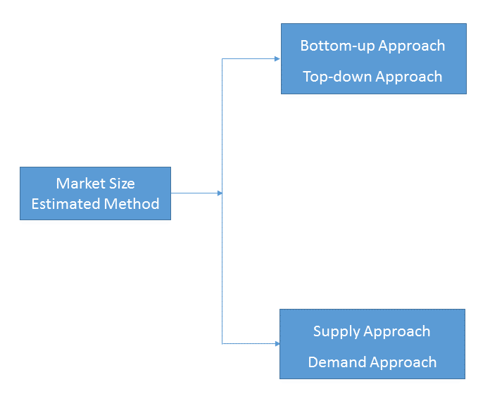
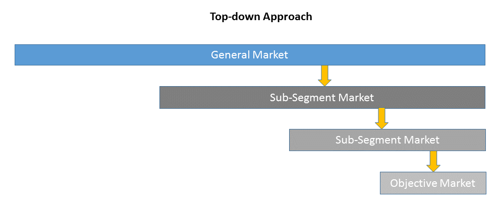
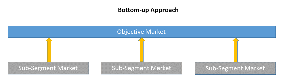
1)Top-down & Bottom-up Approach
Top-down approach uses a general market size figure and determines the percentage that the objective market represents.
Bottom-up approach size the objective market by collecting the sub-segment information.
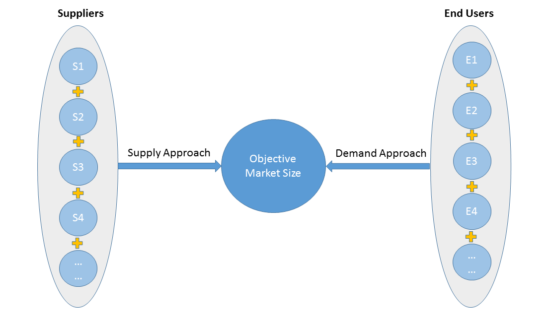
2)Supply & Demand Approach
Supply approach is based on assessments of the size of each competitor supplying the objective market.
Demand approach combine end-user data within a market to estimate the objective market size. It is sometimes referred to as bottom-up approach.
- Forecasting Methodology
- Numerous factors impacting the market trend are considered for forecast model:
- New technology and application in the future;
- New project planned/under contraction;
- Global and regional underlying economic growth;
- Threatens of substitute products;
- Industry expert opinion;
- Policy and Society implication.
- Analysis Tools
1)PEST Analysis
PEST Analysis is a simple and widely used tool that helps our client analyze the Political, Economic, Socio-Cultural, and Technological changes in their business environment.
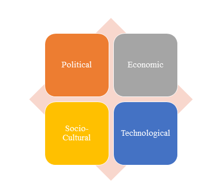
- Benefits of a PEST analysis:
- It helps you to spot business opportunities, and it gives you advanced warning of significant threats.
- It reveals the direction of change within your business environment. This helps you shape what you’re doing, so that you work with change, rather than against it.
- It helps you avoid starting projects that are likely to fail, for reasons beyond your control.
- It can help you break free of unconscious assumptions when you enter a new country, region, or market; because it helps you develop an objective view of this new environment.
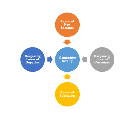
2)Porter’s Five Force Model Analysis
The Porter’s Five Force Model is a tool that can be used to analyze the opportunities and overall competitive advantage. The five forces that can assist in determining the competitive intensity and potential attractiveness within a specific area.
- Threat of New Entrants: Profitable industries that yield high returns will attract new firms.
- Threat of Substitutes: A substitute product uses a different technology to try to solve the same economic need.
- Bargaining Power of Customers: the ability of customers to put the firm under pressure, which also affects the customer's sensitivity to price changes.
- Bargaining Power of Suppliers: Suppliers of raw materials, components, labor, and services (such as expertise) to the firm can be a source of power over the firm when there are few substitutes.
- Competitive Rivalry: For most industries the intensity of competitive rivalry is the major determinant of the competitiveness of the industry.
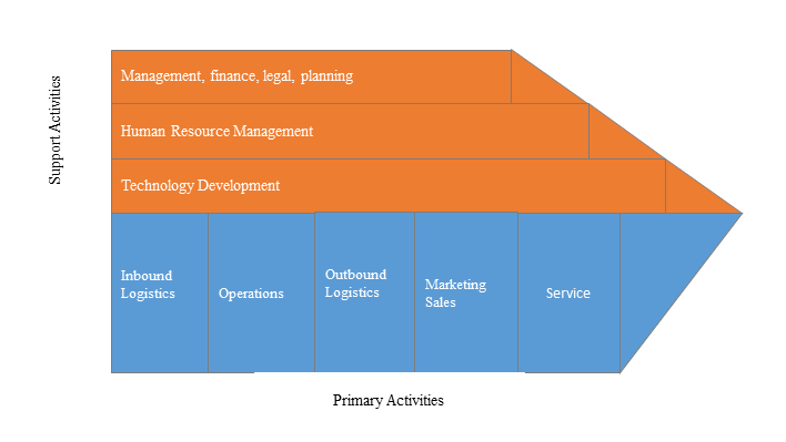
3)Value Chain Analysis
Value chain analysis is a tool to identify activities, within and around the firm and relating these activities to an assessment of competitive strength. Value chain can be analyzed by primary activities and supportive activities. Primary activities include: inbound logistics, operations, outbound logistics, marketing & sales, service. Support activities include: technology development, human resource management, management, finance, legal, planning.
4)SWOT Analysis
SWOT analysis is a tool used to evaluate a company's competitive position by identifying its strengths, weaknesses, opportunities and threats. The strengths and weakness is the inner factor; the opportunities and threats are the external factor. By analyzing the inner and external factors, the analysis can provide the detail information of the position of a player and the characteristics of the industry.
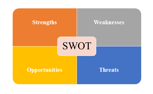
- Strengths describe what the player excels at and separates it from the competition
- Weaknesses stop the player from performing at its optimum level.
- Opportunities refer to favorable external factors that the player can use to give it a competitive advantage.
- Threats refer to factors that have the potential to harm the player.
- Data Sources
| Primary Sources | Secondary Sources |
|---|---|
| Face to face/Phone Interviews with market participants, such as: Manufactures; Distributors; End-users; Experts. Online Survey |
Government/International Organization Data: Annual Report/Presentation/Fact Book Internet Source Information Industry Association Data Free/Purchased Database Market Research Report Book/Journal/News |