Confocal Laser Endomicroscopy Market Analysis 2026-2031
- Single User License (1 Users) $ 3,500
- Team License (2~5 Users) $ 4,500
- Corporate License (>5 Users) $ 5,500
The global Confocal Laser Endomicroscopy (CLE) market size will reach between 30 million to 50 million USD in 2026, and the market is projected to expand at a compound annual growth rate (CAGR) of 7.5% to 12.5% through 2031. This expansion is governed by an escalating clinical imperative: bypassing the delays, procedural morbidity, and diagnostic blind spots inherent to conventional excisional biopsies. Operating at magnifications reaching 1,000 times conventional macroscopic endoscopy, CLE integrates miniaturized laser scanning architectures into flexible catheters, rigid laparoscopic guides, and needle-based interventional vectors. By generating optical sections at lateral depths down to 70 micrometers beneath the tissue surface at frame rates of 8 to 12 frames per second, the technology delivers instant cellular assessment across human clinical settings and companion animal diagnostics.
Technical Operating Principle
The biomechanical efficacy of CLE depends on selective optical spatial filtering combined with dedicated laser fluorescence excitation:
* Laser Light Excitation and Optical Delivery: A console-housed coherent light engine—primarily operating at a 488-nanometer wavelength for blue-laser-induced fluorescein visualization, or 785 nanometers for near-infrared molecular fluorophores—emits focused excitation beams. These photons are directed into high-density coherent imaging bundles comprising tens of thousands of individually drawn optical fiber cores packaged within flexible catheters possessing outer diameters as small as 1 millimeter.
* Subsurface Depth Scanning and Spatial Filtering: The emitted laser beam addresses either topically applied contrast reagents, intravenous sodium fluorescein, or endogenous fluorophores. The targeted tissue emits longer-wavelength fluorescence. Reflected light and out-of-focus background photons are directed through an integrated distal micro-objective lens assembly and back through an internal spatial pinhole aperture. This physical pinhole rejects out-of-focus light rays, ensuring that only signals from the calibrated focal plane (typically positioned 0 to 70 micrometers beneath the epithelial surface) are registered by the photodetector.
* Signal Reconstruction and Video Mosaicing: Emitted signal intensities from individual fiber cores are continuously processed through proprietary algorithmic engines. Digital signal processors deploy motion-compensation filters, frame-freeze triggers, and dynamic spatial mosaicing to correct for physiological peristalsis and respiratory motion artifacts. The output provides high-fidelity, real-time digital reconstructions that directly correlate with conventional fixed-tissue Hematoxylin and Eosin (H&E) paraffin sections.
Core Clinical Value Proposition
CLE shifts the interventional diagnostic paradigm from unguided physical biopsy sampling to targeted, in vivo optical characterization:
* Mitigation of Sampling Artifacts and False Negatives: Conventional macroscopic mucosal sampling is fundamentally vulnerable to geographic misplacement. By contrast, probe-based CLE (pCLE) enables comprehensive surface scanning of suspect mucosa prior to mechanical intervention, reducing the false-negative rates associated with precancerous surveillance regimes.
* Negative Predictive Validation: Clinical surveillance registries consistently confirm a Negative Predictive Value (NPV) reaching 98% in surveillance protocols such as Barrett's esophagus. This metric gives endoscopists the operational confidence to defer non-essential physical tissue excision, diminishing bleeding risks, perforation incidents, and procedural costs.
* Immediate Intraoperative Triage: By generating immediate microstructural insights, CLE eliminates multi-day laboratory processing intervals for frozen sections, empowering clinicians to finalize endoscopic mucosal resections, verify tumor-free margins during organ-sparing oncology surgeries, and prevent unnecessary major surgical operations for indolent cystic lesions.
Downstream Applications and Market Demand
● Human Clinical Healthcare Market
Human clinical healthcare constitutes the principal demand source, driven by six clinical disciplines:
* Gastroenterology and Pancreaticobiliary Systems: The highest-volume clinical application. Within Barrett's Esophagus (BE) and gastroesophageal reflux disease surveillance, pCLE elevates neoplasia detection rates by 243% compared to conventional four-quadrant Seattle biopsy protocols, demonstrating 97% sensitivity and 98% NPV. For Pancreatic Cystic Lesions, where traditional endoscopic ultrasound fine-needle aspiration (EUS-FNA) cytologic yields fail to diagnose greater than 50% of presentations, needle-based CLE (nCLE) provides direct intra-cystic visualization. This yields 100% specificity for benign serous cystadenomas and 99% accuracy for mucinous pre-malignant cysts, generating an estimated health-economic savings of 4,757 USD per patient in avoidable surgical operations. In indeterminate biliary strictures, probe passage through endoscopic retrograde cholangiopancreatography (ERCP) yields an 88% sensitivity for cholangiocarcinoma detection, outpacing conventional brush cytology (54% to 59%). In lower gastrointestinal functional disorders, confocal endomicroscopy dynamically visualizes mucosal barrier breakdown and fluorescein extravasation following luminal allergen challenge, predicting positive responses to targeted exclusion diets in 83% of evaluated patients.
* Interventional Pulmonology and Robotic Integration: Solves the localized diagnostic yield limitation observed in robotic bronchoscopy. While shape-sensing robotic platforms navigate deep into the peripheral pulmonary segments, tissue confirmation remains problematic. Ultra-fine nCLE probes (such as the AQ-Flex 19) advanced through robotic aspiration needles confirm a "Tool-in-Lesion" verification in 95% of deployments, compared to roughly 45% for rapid on-site cytologic evaluation (ROSE), securing a 94% definitive diagnostic confirmation rate for peripheral lung malignancies across a high-volume interventional market.
* Urology and Endourology: Directly targets non-muscle-invasive bladder cancer, which exhibits recurrence rates approaching 75%. pCLE allows endourologists to perform microscopic evaluation of the urothelial lining via standard cystoscopic and ureteroscopic channels, securing 100% diagnostic accuracy for high-grade intraepithelial dysplasia and demarcating upper urinary tract transitional cell carcinoma margins.
* Surgical Oncology and Molecular Margin Demarcation: Handheld and rigid confocal miniprobes deliver real-time surgical margin assessments during breast-conserving resections and head-and-neck oncological procedures. Operating platforms configured for near-infrared wavelengths (such as 785 nanometers) detect molecularly targeted fluorophore tracers administered systemically prior to surgery, identifying microscopic residual nests within the tumor bed.
* Point-of-Care Slide-Free Digital Pathology: Tabletop and compact point-of-care CLE scanners allow the instantaneous imaging of gross surgical specimens without the need for mechanical slicing, tissue freezing, or slide fixation. Digital cellular datasets are automatically routed across hospital local area networks directly to off-site pathology specialists for telepathology evaluations.
* Artificial Intelligence and Algorithmic Clinical Guidance: Deep-learning neural networks integrated directly into CLE platform consoles analyze continuous confocal video streams in real time. These algorithms flag cellular atypia, architectural disorganization, and microvascular leakage, reducing inter-observer discordance and flattening the clinical learning curve for non-academic community hospital deployments.
● Veterinary and Companion Animal Oncology Market
The non-human veterinary sector has transformed from an experimental field into an active commercial vector:
* Companion Animal Neoplasia and Surgical Oncology: Dedicated veterinary confocal systems, exemplified by Optiscan's InSpecta platform unveiled in mid-2025, facilitate immediate cellular evaluation across canine, feline, and equine oncological surgeries. Real-time assessment of soft tissue sarcoma margins, mast cell tumors, and oral melanomas mitigates reliance on outsourced multi-day veterinary histology services.
* Translational Research and Preclinical Drug Development: Highly portable fluorescence micro-endoscopy engines (such as the ViewnVivo platform) are deployed throughout global pharmaceutical research institutes and academic laboratories, supporting longitudinal in vivo drug target engagement, cellular dynamic tracking, and microvascular permeability assessments in living small-animal models without requiring interval animal sacrifice.
Regional Market Dynamics
● North America
North America acts as the primary revenue generator for commercial CLE platforms. The regional market benefits from approximately 14,700 high-capacity endoscopy units across the United States, with initial commercial efforts prioritizing approximately 1,100 high-throughput tertiary gastroenterology centers and Ambulatory Surgical Centers (ASCs). Clinical demand is sustained by high disease prevalence, including 3.6 million adults diagnosed with Barrett's esophagus, pervasive GERD manifestations, and half a million annual diagnostic bronchoscopy procedures.
The regional market is supported by four specialized Current Procedural Terminology (CPT) billing codes (Category I codes 43206, 43252, 88375, and Category III code 0397T), which facilitate facility and physician professional fee reimbursement under the Medicare Outpatient Prospective Payment System (OPPS) and Ambulatory Payment Classifications (APC). Procedural adoption is endorsed within practice guidelines published by the American Gastroenterological Association (AGA) and the American Society of General Surgeons (ASGS). Regional commercial scaling is reinforced by strategic marketing alliances, including distribution pacts linking nCLE consoles with therapeutic endoscopic ultrasound product suites.
● Asia-Pacific
Asia-Pacific represents the fastest-expanding territory by procedure volume, supported by an aggregate operational base exceeding 30,000 endoscopy suites across mainland China and Japan. In China, market penetration strategies focus on tier-one tertiary hospitals out of an institutional universe of more than 23,000 facilities. The regional operating climate underwent a notable transition following the late-2025 National Medical Products Administration (NMPA) Class III registration for third-generation pCLE platforms, clearing paths for domestic commercial distribution following the termination and restructuring of legacy joint venture frameworks.
In Japan, commercialization focuses on 200 to 300 academic university hospitals via the Pharmaceuticals and Medical Devices Agency (PMDA) regulatory pathway. Australia functions as an optical engineering center and a primary surgical trial jurisdiction, sustained by federal research initiatives, including R&D Tax Incentive programs and CRC-P translational frameworks. South Korea provides established reimbursement tracking for confocal mucosal assessments, while regional equipment adoption across Taiwan, China, and Southeast Asian facilities continues to track higher gastrointestinal cancer screening volumes.
● Europe
Europe maintains a well-established clinical footprint across roughly 15,000 hospital endoscopy departments. Commercial expansion across member states has been dictated by compliance with the European Medical Device Regulation (MDR 2017/745), a transition requiring comprehensive clinical data updates that key console lines and companion flexible probes (such as GastroFlex, ColoFlex, CholangioFlex, and AQ-Flex 19) achieved post-2025.
Reimbursement mechanisms remain nationally fragmented. In France, endomicroscopic interventions are cataloged under the Common Classification of Medical Procedures (CCAM) with clear compensation for gastroenterologists and anesthesiologists; public health bodies (such as Haute Autorité de Santé) consistently re-evaluate nCLE health-economic utility alongside updated guidelines from the European Society of Gastrointestinal Endoscopy (ESGE). In Germany, multi-site private hospital networks (such as the HELIOS group) utilize activity-based G-DRG allocations to finance probe utilization, particularly for functional food intolerance diagnostics and pancreaticobiliary strictures. Channel relationships drive ongoing market adoption across the United Kingdom, Switzerland, and Italy.
● Latin America
Commercial deployments across Latin America rely primarily on exclusive third-party specialty distributors responsible for handling local sanitary registrations, importation customs, and clinical training programs. In Ecuador, long-term regulatory clearances cover confocal consoles and miniprobes through late 2027. Commercialization across private and academic institutions in Brazil, Mexico, and Colombia targets specialized centers handling esophageal cancer screenings, pancreatic diagnostic evaluations, and food-induced barrier dysfunction testing.
● Middle East and Africa (MEA)
The MEA territory represents an emerging market segment governed by capital equipment sales to tertiary referral facilities, premier military health organizations, and specialized academic institutions. High-growth areas center on the United Arab Emirates, Saudi Arabia, and select private healthcare hubs in South Africa. Vendor strategies rely on regional distributor networks with specialized divisions in advanced therapeutic endoscopy and interventional pulmonology.
Supply Chain and Value Chain Architecture
● Upstream Component and Raw Material Specialization
The CLE manufacturing supply chain is exposed to severe supplier concentration risks, driven by the specialized physics of miniature optical components:
* Ultra-High-Density Coherent Fiber Bundles: The central structural bottleneck of the entire sector. A pCLE probe requires up to tens of thousands of individual, sub-micron optical fiber cores fused into a flexible bundle with an outer diameter under 1 millimeter, maintaining uniform light transmission with minimal cross-talk. Global commercial manufacturing is largely dependent on single-source Japanese optics specialists, primarily the Fujikura Group and its Fibertech division. Mitigation strategies deployed by device manufacturers rely on rigorous master supply agreements incorporating rolling multi-year firm purchase orders, extended six-month minimum termination clauses, and safety buffer inventories designed to deliver up to five times historical annual volumes in the event of manufacturing interruptions.
* Optoelectronic Sub-Assemblies: Comprises solid-state diode lasers calibrated precisely to single-line emissions (488 nm or 785 nm), micro-electromechanical systems (MEMS) scanning actuators, chromatic beam splitters, and custom anti-reflective micro-objective lenses. Sourcing relies on a limited cohort of specialized industrial precision optics manufacturers distributed across Japan, Germany, and the United States.
● Midstream Assembly, Fabrication, and IP Defensibility
Value capture within the midstream tier is concentrated in proprietary optical-mechanical-software integration, prompting manufacturers to maintain an asymmetric, hybrid operational profile:
* Proprietary In-House Integration: Console vendors deliberately keep high-margin processes in-house. This includes high-precision optical fiber alignment, micro-lens spatial bonding, scanning driver programming, and dynamic image-processing software configuration. Conversely, non-proprietary mechanical framing, sheet-metal console stamping, standard electronic printed circuit boards (PCBs), and medical-grade power modules are outsourced to contract manufacturers.
* Quality Management and Regulatory Moats: Manufacturing infrastructure operates under ISO 13485 certification, FDA Quality System Regulation (21 CFR Part 820), and EU MDR Annex IX audits. Transitioning an optical fiber supplier requires comprehensive console re-benchmarking, biocompatibility re-testing, sterilization re-validation, and formal regulatory submissions, presenting a high switching barrier.
* Intellectual Property Portfolios: Market incumbents deploy defensive patent strategies covering resonant vibrating fiber scanners, needle-compatible catheter housings, video frame-mosaicing algorithms, real-time background subtraction methods, and multi-wavelength laser coupling modules. Leading portfolios often encompass between 250 and 300 active patents globally, creating substantial technical barriers for emerging market entrants.
● Downstream Delivery and Clinical Workflow Integration
The downstream ecosystem connects high-complexity clinical departments with centralized hospital informatics:
* Institutional Deployment Hubs: Systems are installed across academic university medical complexes, regional tertiary referral centers, high-volume outpatient ASCs, and specialized companion animal surgical hospitals.
* Enterprise Imaging and Interoperability: Modern CLE platforms are designed to export DICOM-compliant video streams directly into institutional Picture Archiving and Communication Systems (PACS) and cloud-linked telepathology repositories. This facilitates real-time remote consultations between the surgical suite, the endoscopy theater, and off-site pathology specialists.
Company Profiles and Competitive Moats
● Mauna Kea Technologies: Founded and headquartered in Paris, France, the company functions as the principal pioneer and developer of probe-based Confocal Laser Endomicroscopy under the Cellvizio brand.
* Operational Architecture and Strategic Focus: Operates console architectures based on the Cellvizio 100 Series and the Cellvizio I.V.E., utilizing 488 nm and 785 nm excitation sources paired with an extensive suite of single-use and reprocessable Confocal Miniprobes. Strategic initiatives focus on securing high-margin recurring consumable probe usage across four clinical sub-segments: upper and lower gastroenterology (GastroFlex, ColoFlex), cholangiopancreatoscopy (CholangioFlex), robotic interventional pulmonology (AlveoFlex, BronchoFlex), and fine-needle aspiration pancreatic cysts (AQ-Flex 19).
* Operational Moats: Unmatched intellectual property portfolio encompassing more than 265 issued patents worldwide, established CPT Category I and III coding infrastructure in the United States, CE mark under EU MDR, and NMPA registration in China. Maintains direct clinical-distribution alignments with interventional medical device conglomerates.
● Optiscan Imaging Ltd: An Australian optical engineering company based in Melbourne, specializing in the design and miniaturization of confocal laser scanning microscopy systems.
* Operational Architecture and Strategic Focus: Built upon proprietary single-fiber architecture where a single vibrating optical fiber functions simultaneously as the point source, the scanning actuator, and the pinhole detector. The product suite includes InVivage (a handheld system tailored for oral cancer screening and margin demarcation), ViewnVivo (preclinical live small-animal translational imaging), and InSpecta (unveiled in mid-2025 as a dedicated veterinary clinical endomicroscope). Concurrently provides core scanning hardware engines for third-party medical technology partners.
* Operational Moats: Proprietary mechanical fiber-vibrating scanner intellectual property that eliminates bulky internal mirrors and beam-splitting prisms; extensive research affiliations with academic veterinary and surgical oncology research institutions; recipient of sustained Australian translational research grant funding.
● Carl Zeiss Meditec: A global medical technology company operating as the optical and surgical visualization division of the Carl Zeiss Group, headquartered in Jena, Germany.
* Operational Architecture and Strategic Focus: Integrates confocal endomicroscopy into surgical operating environments via the ZEISS CONVIVO platform, an intraoperative digital cellular visualization system developed in collaboration with Optiscan. The system allows handheld real-time histological assessment of brain tumors and neurosurgical resection boundaries using sodium fluorescein.
* Operational Moats: Seamless integration across surgical visualization systems (such as the KINEVO 900 platform); direct market access to neurosurgical operating departments globally; proprietary enterprise digital telepathology workflow software enabling instantaneous surgical-pathological consultations during active cranial procedures.
● Viestar Medical Technology Co. Ltd. (Jingwei Shida): A domestic optical biopsy enterprise based in China, recognized as an early domestic developer to clear Class III medical device regulatory thresholds for probe-based confocal systems.
* Operational Architecture and Strategic Focus: Commercializes the BriteView confocal platform. Focuses on producing sub-millimeter flexible optical micro-probes compatible with standard 2.8 mm or smaller biopsy channels of standard gastrointestinal and respiratory endoscopes. Operates proprietary Unif background-scattering suppression and image-filtering algorithms.
* Operational Moats: Holds a Class III registration from China's NMPA, shielding the platform from foreign device displacement mandates in domestic public tertiary hospitals; domestic cost advantages in laser console housing fabrication; direct distribution across Chinese provincial hospital procurement networks.
● Hisky Medical Emerging from clinical acoustic and optical engineering programs at Tsinghua University, China, the company expanded beyond its foundational liver diagnostic portfolio (FibroTouch) into multi-specialty cellular endomicroscopy.
* Operational Architecture and Strategic Focus: Manufactures the CelTouch (BrightP and BrightX series) probe-based endomicroscopy platforms. Engineered to serve as a cellular-level, multi-departmental optical biopsy solution compatible with standard flexible endoscopic instrumentation across gastroenterology, interventional pulmonology, and urology.
* Operational Moats: Integrated production and R&D lines; machine-learning signal processing modules optimized for deep mucosal penetration; broad commercial reach stemming from an established institutional sales footprint across national hospital liver centers.
Strategic Opportunities and Structural Challenges
● Strategic Growth Catalysts
* Robotic Platform Embedment and "Tool-in-Lesion" Verification: As lung cancer management migrates toward early-stage peripheral nodule resections, robotic bronchoscopy platforms face a common operational limitation: reaching a sub-solid peripheral lesion does not guarantee accurate diagnostic needle localization. The deployment of ultra-flexible nCLE miniprobes down the core of interventional aspiration needles confirms cellular-level malignant localization prior to biopsy execution. This real-time validation addresses diagnostic yield gaps, positioning endomicroscopic verification as an integral guidance mechanism for the surgical robotics sector.
* Expansion into Companion Animal Clinical Oncology: Veterinary medicine represents a commercial pathway largely unencumbered by human public payer reimbursement restrictions and multi-year centralized regulatory trials. With companion animal owners increasingly electing advanced oncological resections, dedicated veterinary confocal engines (e.g., InSpecta) bridge the gap for private specialty animal hospitals that require real-time margin clearance without waiting days for off-site pathology reports.
* Point-of-Care Slide-Free Specimen Scanners: Moving beyond catheter-based luminal endoscopy, adapting confocal engines into standalone, benchtop pathology tools presents a viable growth trajectory. These platforms scan fresh, non-excised surgical specimens directly within operating suites, generating virtual digital histology sections that preserve original tissues for subsequent molecular testing and genomic profiling.
● Critical Market Inhibitors and Structural Bottlenecks
* Institutional Reimbursement Fragility and Payer Coding Volatility: The long-term commercial trajectory of CLE remains linked to procedural reimbursement stability. In the United States, prior shifts in Centers for Medicare & Medicaid Services (CMS) hospital outpatient reimbursement schedules—often precipitated by inconsistent cost reporting by individual hospital billing departments—produced volatility in Pay-Per-Use (PPU) probe revenues. The lack of standardized, high-rate physician fee schedules across international public health programs (such as select European frameworks and Asian health systems) restricts platform placements to well-funded academic departments, slowing community adoption.
* Stringent Regulatory Hurdles Under MDR and International Agencies: The transition to the European Medical Device Regulation (MDR) has increased operational costs across the sector. Demonstrating long-term clinical safety equivalence, updating post-market surveillance systems, and clearing clinical evaluations require significant capital expenditure, extending commercial launch timelines for next-generation consoles and disposable probe designs.
* Severe Upstream Fiber Bundle Monopolization: The single-source reliance on specialized Japanese manufacturers for ultra-high-density coherent optical fiber bundles represents a notable supply chain vulnerability. Because probe performance depends directly on the core counts and flexible properties of these proprietary conduits, any manufacturing downtime, geopolitical trade bottleneck, or material scarcity at the sub-supplier tier leaves the entire market susceptible to production delays and margin compression. Strategic inventory stockpiling and defensive long-term frame purchasing agreements remain the only viable mitigations available to device manufacturers today.
1.1 Executive Synthesis & High-Level Market Perimeter 1
1.2 Multi-Tiered Research Methodology & Quantitative Triangulation Models 3
1.3 Primary Source Matrix, Expert Delphi Panels & Key Secondary Corroborations 4
1.4 Macroeconomic Baselines, Currency Parity & Forecast Calibration Hypotheses 5
1.5 Definitional Standards, Nomenclature & Analytical Abbreviations 6
Chapter 2 Global CLE Market Dynamics & Structural Trajectory (2021-2031) 7
2.1 Global Market Valuation, Longitudinal Revenue Run-Rates and Volume Evolution 7
2.2 Macro Secular Drivers: In Vivo Real-Time Optical Biopsy and Diagnostic Paradigm Shifts 8
2.3 Structural Headwinds, Capital Cost Bottlenecks and Pathologist Learning Curves 9
2.4 Cross-Disciplinary Convergence: Artificial Intelligence, Deep Learning and Automated Histological Grading 10
2.5 Price Erosion Curves, Probe Reusability Cycles and Total Cost of Ownership Modeling 11
2.6 Competitive Concentration Index, Herfindahl-Hirschman Assessment and Industry Consolidation 12
Chapter 3 Upstream Supply Chain Architecture & Advanced Component Ecosystem 13
3.1 Upstream Value Chain Stratification and Critical Raw Material Interdependencies 13
3.2 Solid-State & Fiber Laser Sources: Laser Diode Dynamics (488 nm, 660 nm, Near-Infrared Modules) 14
3.3 Micro-Optical Assemblies: Miniature Objective Lenses, MEMS Scanners and Scanning Mirrors 15
3.4 Coherent Fiber Optic Bundles: Fiber Drawing Precision, Core Densities and Transmission Attenuation 16
3.5 Contrast Agents and Fluorophore Sourcing Dynamics: Fluorescein Sodium, Acriflavine, Cresyl Violet 17
3.6 Supply Chain Vulnerability Matrix, Dual-Sourcing Strategies and Geopolitical Chokepoints 18
Chapter 4 Global Confocal Laser Endomicroscopy Market by Architecture & Product Modality 19
4.1 Probe-based Confocal Laser Endomicroscopy (pCLE) Systems 19
4.1.1 Optical Conduit Architecture, Catheter Working Channel Compatibility and Maneuverability 19
4.1.2 Global Revenue, Unit Deployments and Annual Fleet Growth (2021-2031) 20
4.2 Needle-based Confocal Laser Endomicroscopy (nCLE) Systems 21
4.2.1 Fine Needle Aspiration/Biopsy (EUS-FNA/FNB) Integration Mechanics 21
4.2.2 Global Revenue, Adoption Vectors and Market Penetration Curves (2021-2031) 22
4.3 Integrated / Endoscope-based Confocal Laser Endomicroscopy (eCLE) Systems 23
4.3.1 Built-in Distal Tip Miniature Scanning Assemblies and Optical System Synergy 23
4.3.2 Revenue Performance, Capital Replacement Cycles and Obsolescence Metrics (2021-2031) 24
Chapter 5 Global CLE Market Breakdown by Application Vertical 25
5.1 Human Clinical Healthcare Ecosystem 25
5.1.1 Gastroenterology: Barrett's Esophagus, Gastric Precancerous Lesions, Colorectal Polyps and Pancreatic Cysts 25
5.1.2 Pulmonology: Peripheral Pulmonary Nodules, Airway Epithelium and Alveolar Microstructures 26
5.1.3 Urology & Gynecology: Non-Muscle Invasive Bladder Cancer (NMIBC) and Cervical Epithelial Dysplasia 27
5.1.4 Surgical Oncology & Intraoperative Tissue Guidance: Brain Neoplasms and Head & Neck Margins 28
5.2 Veterinary & Pet Healthcare Ecosystem 29
5.2.1 Companion Animal Oncology: Canine and Feline Soft Tissue Margins and Neoplastic Lesions 29
5.2.2 Veterinary Translational Research, Exotic Animal Diagnostics and Equine Sports Medicine 30
Chapter 6 Global Trade Flow Mechanics, Regulatory Milestones & Reimbursement Frameworks 31
6.1 Cross-Border Logistics, Specialized Optical Assembly Shipping and Export Compliance 31
6.2 Global Trade Corridors: High-Precision Component Flows between North America, Europe and East Asia 32
6.3 Regulatory Gateways: FDA 510(k)/PMA, EU MDR Class IIa/IIb Reclassification and NMPA Fast-Track Tracks 33
6.4 Reimbursement Dynamics: CPT Category I/III Coding Infrastructure, DRG Mapping and Outpatient Prospective Payment Coverage 34
6.5 Cross-Regional Health Technology Assessment (HTA) Adoption Hurdles and Clinical Efficacy Proofs 35
Chapter 7 North American Confocal Laser Endomicroscopy Market Landscape 36
7.1 Regional Market Sizing, Installed Base Velocity and Clinical Utilization Rates (2021-2031) 36
7.2 United States: Tier-1 Academic Medical Centers, ASC Penetration and Capital Budgeting Dynamics 37
7.3 Canada: Provincial Procurement Consortiums, Special Access Programs and Clinical Trials Footprint 40
Chapter 8 European Confocal Laser Endomicroscopy Market Landscape 42
8.1 European Market Overview, Cross-Border Tender Structures and MDR Transition Pressures (2021-2031) 42
8.2 Germany: Fraunhofer Research Influence, Optical Cluster Hubs and Inpatient Hospital Funding 43
8.3 France: Early Technological Incubation, University Hospital Clinical Trials and National Health Insurance Inroads 45
8.4 United Kingdom: NHS Innovation Procurement Frameworks, NICE Diagnostic Guidance and Veterinary Networks 46
8.5 Rest of Europe Market 47
Chapter 9 Asia-Pacific Confocal Laser Endomicroscopy Market Landscape 49
9.1 Asia-Pacific Macro Sizing, Healthcare Modernization and Leapfrogging Potential (2021-2031) 49
9.2 China: Domestic Substitution Policies, High-Volume Class 3A Hospitals and Fast-Growing Animal Clinic Chains 50
9.3 Japan: Endoscopic Innovation Foundations, PMDA Approval Pathways and Clinical Workflow Integration 53
9.4 Australia: Precision Medical Device Clusters, Clinical Translation Frameworks and Outbound Trade 54
Chapter 10 Rest of the World Confocal Laser Endomicroscopy Market Footprint 56
10.1 Latin America: Brazil and Mexico Clinical Adoption, Import Tariffs and Tertiary Private Centers 56
10.2 Middle East & Africa: GCC Premium Healthcare Infrastructure, Medical Tourism Hubs and Public Tender Models 58
Chapter 11 Competitive Benchmarking & Corporate Intelligence: Tier-1 Market Players 60
11.1 Carl Zeiss Meditec 60
11.1.1 Corporate Structure, Global Footprint and Advanced Optical Imaging Portfolio Overview 60
11.1.2 Product-Level Strategic Positioning: System Integration and Intraoperative Visualization 61
11.1.3 Strategic SWOT Matrix (Strengths, Weaknesses, Opportunities, Threats) 62
11.1.4 Financial Performance: Revenue, Direct Manufacturing Costs, Gross Margin and Market Share (2021-2026) 63
11.2 Optiscan Imaging Ltd 64
11.2.1 Corporate Genesis, Miniature Scanning Architecture and Global Commercialization Agreements 64
11.2.2 Clinical and Translational Product Array: Digital Endomicroscopy Hardware & Software Solutions 65
11.2.3 Strategic SWOT Matrix (Strengths, Weaknesses, Opportunities, Threats) 66
11.2.4 Financial Performance: Revenue, Direct Manufacturing Costs, Gross Margin and Market Share (2021-2026) 67
11.3 Mauna Kea Technologies 68
11.3.1 Enterprise Background, Pioneer Proprietary Cellvizio Platform Architecture and Clinical Depth 68
11.3.2 Human Clinical Healthcare and Preclinical Veterinary/Translational Diagnostic Offerings 69
11.3.3 Strategic SWOT Matrix (Strengths, Weaknesses, Opportunities, Threats) 70
11.3.4 Financial Performance: Revenue, Direct Manufacturing Costs, Gross Margin and Market Share (2021-2026) 71
11.4 Viestar Medical Technology Co. Ltd. 72
11.4.1 Corporate Foundation, Domestic Market Position in China and R&D Capabilities 72
11.4.2 Probe-based and Needle-based Clinical Imaging Solutions Portfolio 73
11.4.3 Strategic SWOT Matrix (Strengths, Weaknesses, Opportunities, Threats) 74
11.4.4 Financial Performance: Revenue, Direct Manufacturing Costs, Gross Margin and Market Share (2021-2026) 75
11.5 Hisky Medical 76
11.5.1 Enterprise Architecture, Multi-Modality Imaging Platforms and Clinical Expansion Strategy 76
11.5.2 CLE Commercial Strategy, Cross-Disciplinary Synergies and Clinical Penetration 77
11.5.3 Strategic SWOT Matrix (Strengths, Weaknesses, Opportunities, Threats) 78
11.5.4 Financial Performance: Revenue, Direct Manufacturing Costs, Gross Margin and Market Share (2021-2026) 79
Chapter 12 Strategic Technological Trajectories & Future Innovation Vectors (2027-2031) 80
12.1 Multi-Photon and Dual-Axis Confocal Architectures: Depth Penetration and Label-Free Autofluorescence 80
12.2 Integration with Robotic Endoluminal Platforms, Bronchoscopy Robotics and Micro-Manipulators 81
12.3 Molecular Imaging Probes, Targeted Fluorophores and Tumor-Specific Endomicroscopy 82
12.4 Generative AI-Assisted Real-Time Histopathological Pattern Segmentation and Semantic Interpretation 83
12.5 Miniaturization Paradigms, Disposable Probe Cost Curves and Decentralized Diagnostic Clinics 84
Table 2 Global Confocal Laser Endomicroscopy Market Revenue and Growth Projections (2021-2031) 7
Table 3 Global Confocal Laser Endomicroscopy Unit Deployments and Average Selling Price (ASP) Trends (2021-2031) 11
Table 4 High-Precision Optical Component Suppliers and Raw Material Sourcing Footprint 14
Table 5 Key Optical Fiber Bundle Suppliers: Technical Parameters, Densities and Commercial Terms 16
Table 6 Global CLE Market Revenue by Architecture (Probe-based, Needle-based, Integrated) (2021-2031) 20
Table 7 Global Probe-based CLE (pCLE) Revenue Breakdown by Major Geographic Region (2021-2031) 20
Table 8 Global Needle-based CLE (nCLE) Revenue Breakdown by Major Geographic Region (2021-2031) 22
Table 9 Global Integrated CLE (eCLE) Revenue Breakdown by Major Geographic Region (2021-2031) 24
Table 10 Global Confocal Laser Endomicroscopy Market Revenue by Application (2021-2031) 25
Table 11 Human Clinical Healthcare CLE Market Revenue by Sub-Specialty (2021-2031) 28
Table 12 Veterinary & Pet Healthcare CLE Market Revenue by Animal Type and Setting (2021-2031) 30
Table 13 Cross-Border Regulatory Approval Roadmaps: FDA, MDR, NMPA and PMDA Comparison 33
Table 14 Existing and Emerging CPT Category Codes and Hospital Outpatient Payment Classifications 34
Table 15 North American CLE Revenue Breakdown by Country (United States, Canada) (2021-2031) 36
Table 16 United States CLE Market Revenue by Architecture Modality (2021-2031) 37
Table 17 United States CLE Market Revenue by Downstream Application Segment (2021-2031) 39
Table 18 European CLE Market Revenue Breakdown by Leading Country (2021-2031) 42
Table 19 Germany CLE Market Revenue by Clinical Application and Hardware Type (2021-2031) 44
Table 20 France CLE Market Revenue by Clinical Application and Hardware Type (2021-2031) 45
Table 21 United Kingdom CLE Market Revenue by Clinical Application and Hardware Type (2021-2031) 46
Table 22 Asia-Pacific CLE Market Revenue Breakdown by Leading Country (2021-2031) 49
Table 23 China CLE Market Revenue and Fleet Size by Product Category (2021-2031) 51
Table 24 Japan CLE Market Revenue and Fleet Size by Product Category (2021-2031) 53
Table 25 Australia CLE Market Revenue and Fleet Size by Product Category (2021-2031) 55
Table 26 Rest of the World CLE Market Revenue Breakdown by Region (2021-2031) 57
Table 27 Carl Zeiss Meditec Confocal Laser Endomicroscopy Revenue, Cost and Gross Margin (2021-2026) 63
Table 28 Optiscan Imaging Ltd Confocal Laser Endomicroscopy Revenue, Cost and Gross Margin (2021-2026) 67
Table 29 Mauna Kea Technologies Confocal Laser Endomicroscopy Revenue, Cost and Gross Margin (2021-2026) 71
Table 30 Viestar Medical Technology Co. Ltd. Confocal Laser Endomicroscopy Revenue, Cost and Gross Margin (2021-2026) 75
Table 31 Hisky Medical Confocal Laser Endomicroscopy Revenue, Cost and Gross Margin (2021-2026) 79
Figure 1 Global Confocal Laser Endomicroscopy Market Sizing and Year-over-Year Trajectory (2021-2031) 8
Figure 2 Confocal Laser Endomicroscopy Value Chain and Intermediary Cost Additions 13
Figure 3 Laser Diode and Scanning Optical Assembly Production Volume Trends 15
Figure 4 Global CLE Market Value Share by Architecture Type (2026 vs. 2031) 19
Figure 5 Global Probe-based vs. Needle-based System Deployment Velocity (2021-2031) 21
Figure 6 Global CLE Market Revenue Share by Application: Human vs. Veterinary Healthcare (2026) 26
Figure 7 Human Clinical Healthcare Segment Penetration Breakdown by Specialty 27
Figure 8 Veterinary & Pet Healthcare CLE Utilization Trajectory (2021-2031) 29
Figure 9 Inter-Regional Supply and Demand Logistics: Key Export Corridors vs. Consumption Sinks 32
Figure 10 North American Installed Base Breakdown by Provider Setting (2021-2031) 37
Figure 11 United States pCLE vs. nCLE Revenue Expansion Curves (2021-2031) 38
Figure 12 European CLE Market Share Distribution by Leading Nation (2026) 43
Figure 13 Germany Healthcare Capital Expenditure on Advanced Diagnostic Endoscopy (2021-2031) 44
Figure 14 Asia-Pacific CLE Annual Growth Rate Outperformance (2021-2031) 50
Figure 15 China Domestic vs. Imported CLE Equipment Market Penetration Shift (2021-2031) 52
Figure 16 Japan High-Definition Endoscopic Optical Imaging Installed Base Dynamics 54
Figure 17 Carl Zeiss Meditec Confocal Laser Endomicroscopy Market Share (2021-2026) 63
Figure 18 Optiscan Imaging Ltd Confocal Laser Endomicroscopy Market Share (2021-2026) 67
Figure 19 Mauna Kea Technologies Confocal Laser Endomicroscopy Market Share (2021-2026) 71
Figure 20 Viestar Medical Technology Co. Ltd. Confocal Laser Endomicroscopy Market Share (2021-2026) 75
Figure 21 Hisky Medical Confocal Laser Endomicroscopy Market Share (2021-2026) 79
Figure 22 Next-Generation Technology Convergence: CLE Integration with Endoscopic Robotic Platforms 82
Research Methodology
- Market Estimated Methodology:
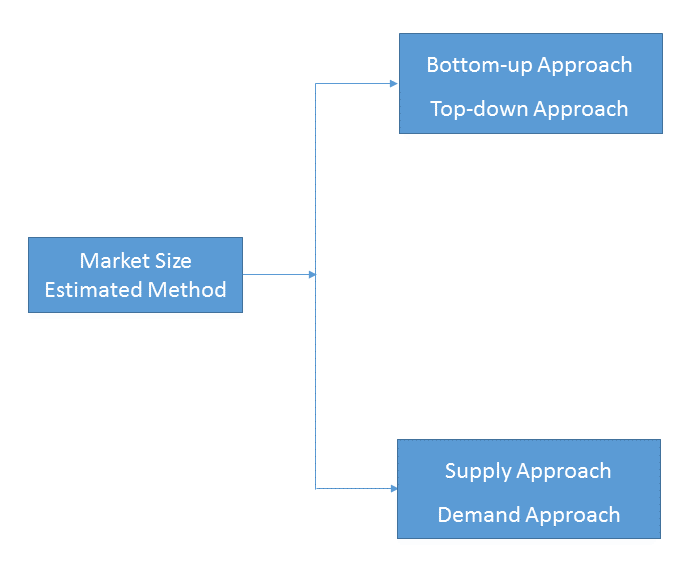
Bottom-up & top-down approach, supply & demand approach are the most important method which is used by HDIN Research to estimate the market size.
1)Top-down & Bottom-up Approach
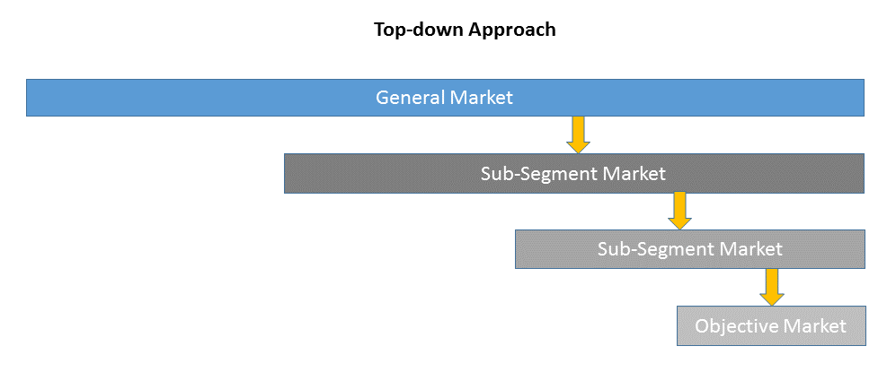
Top-down approach uses a general market size figure and determines the percentage that the objective market represents.
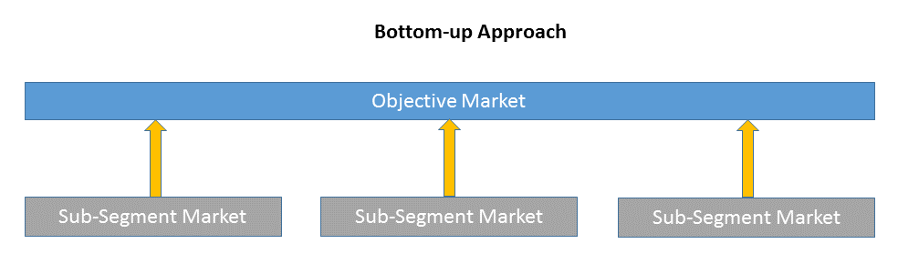
Bottom-up approach size the objective market by collecting the sub-segment information.
2)Supply & Demand Approach
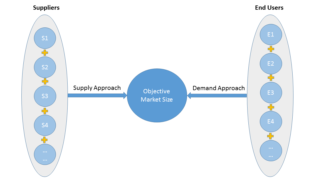
Supply approach is based on assessments of the size of each competitor supplying the objective market.
Demand approach combine end-user data within a market to estimate the objective market size. It is sometimes referred to as bottom-up approach.
- Forecasting Methodology
- Numerous factors impacting the market trend are considered for forecast model:
- New technology and application in the future;
- New project planned/under contraction;
- Global and regional underlying economic growth;
- Threatens of substitute products;
- Industry expert opinion;
- Policy and Society implication.
- Analysis Tools
1)PEST Analysis
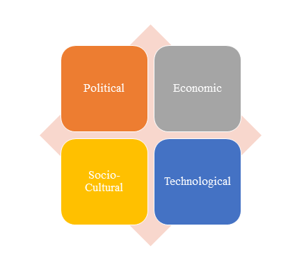
PEST Analysis is a simple and widely used tool that helps our client analyze the Political, Economic, Socio-Cultural, and Technological changes in their business environment.
- Benefits of a PEST analysis:
- It helps you to spot business opportunities, and it gives you advanced warning of significant threats.
- It reveals the direction of change within your business environment. This helps you shape what you’re doing, so that you work with change, rather than against it.
- It helps you avoid starting projects that are likely to fail, for reasons beyond your control.
- It can help you break free of unconscious assumptions when you enter a new country, region, or market; because it helps you develop an objective view of this new environment.
2)Porter’s Five Force Model Analysis
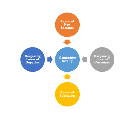
The Porter’s Five Force Model is a tool that can be used to analyze the opportunities and overall competitive advantage. The five forces that can assist in determining the competitive intensity and potential attractiveness within a specific area.
- Threat of New Entrants: Profitable industries that yield high returns will attract new firms.
- Threat of Substitutes: A substitute product uses a different technology to try to solve the same economic need.
- Bargaining Power of Customers: the ability of customers to put the firm under pressure, which also affects the customer's sensitivity to price changes.
- Bargaining Power of Suppliers: Suppliers of raw materials, components, labor, and services (such as expertise) to the firm can be a source of power over the firm when there are few substitutes.
- Competitive Rivalry: For most industries the intensity of competitive rivalry is the major determinant of the competitiveness of the industry.
3)Value Chain Analysis
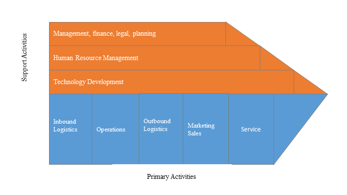
Value chain analysis is a tool to identify activities, within and around the firm and relating these activities to an assessment of competitive strength. Value chain can be analyzed by primary activities and supportive activities. Primary activities include: inbound logistics, operations, outbound logistics, marketing & sales, service. Support activities include: technology development, human resource management, management, finance, legal, planning.
4)SWOT Analysis
SWOT analysis is a tool used to evaluate a company's competitive position by identifying its strengths, weaknesses, opportunities and threats. The strengths and weakness is the inner factor; the opportunities and threats are the external factor. By analyzing the inner and external factors, the analysis can provide the detail information of the position of a player and the characteristics of the industry.
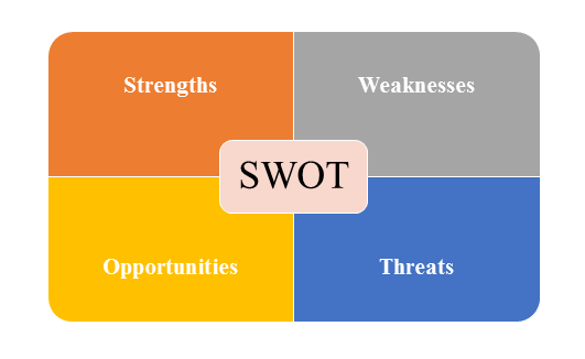
- Strengths describe what the player excels at and separates it from the competition
- Weaknesses stop the player from performing at its optimum level.
- Opportunities refer to favorable external factors that the player can use to give it a competitive advantage.
- Threats refer to factors that have the potential to harm the player.
- Data Sources
| Primary Sources | Secondary Sources |
|---|---|
| Face to face/Phone Interviews with market participants, such as: Manufactures; Distributors; End-users; Experts. Online Survey |
Government/International Organization Data: Annual Report/Presentation/Fact Book Internet Source Information Industry Association Data Free/Purchased Database Market Research Report Book/Journal/News |